


Aperture
The question that haunted the field —
Why do cellular membranes behave like liquid crystals under mechanical stress?
For two decades, membrane biophysics operated under a consensus model that treated lipid bilayers as passive scaffolds. The anomalous stress responses observed in Yoshida et al. (2019) were filed as experimental noise. No one asked why the noise had a pattern.
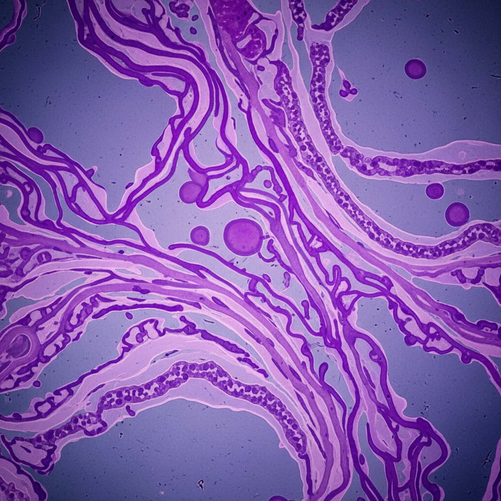
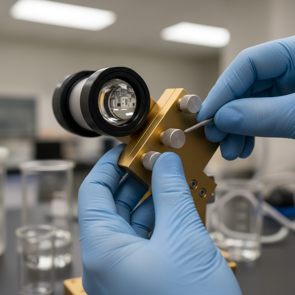
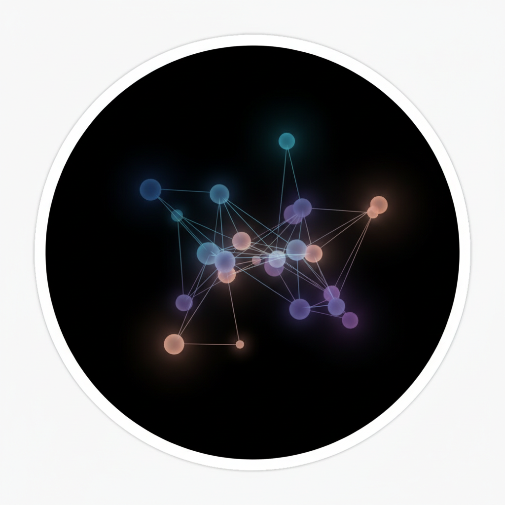
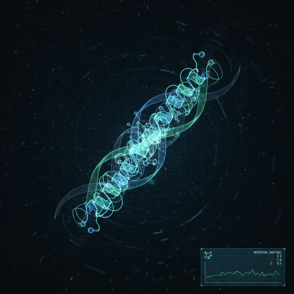
Aperture's Soft Matter group developed a sub-nanometer interferometric lens — a computational-optical hybrid — that could image membrane topology at timescales four orders of magnitude faster than existing cryo-EM approaches. The noise became signal.
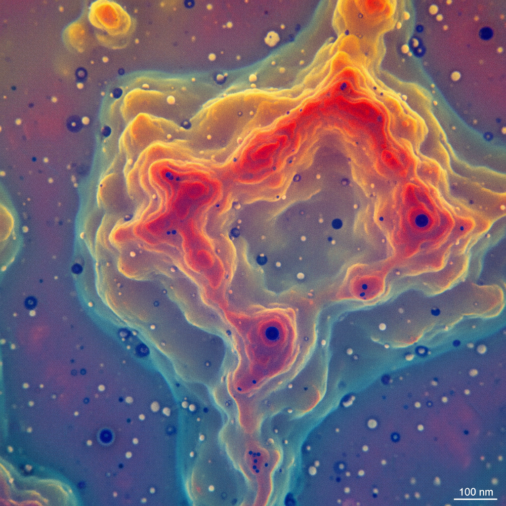
Aperture Interferometric Lens
4,000×
faster than cryo-EM at equivalent resolution
The data that emerged —
The door it opened —
Membrane topology, it turns out, encodes mechanical memory. This finding reframes how we understand cellular ageing, drug delivery barriers, and the structural basis of three previously unexplained genetic disorders. The lens is now being applied to neural tissue.
The question that haunted the field —
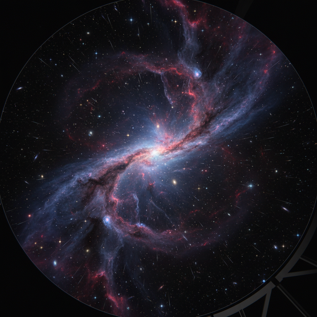
Can spectral residuals in black hole accretion discs map the topology of spacetime curvature?
Standard models treat spectral residuals as instrument artifacts. Aperture's Astrophysics Computation group noticed the residuals correlated — weakly, but consistently — with orbital period anomalies documented in Doeleman et al. (2022). Correlation isn't causation, but it is a question.
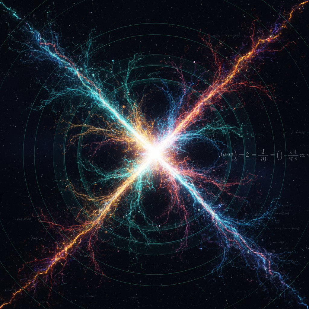
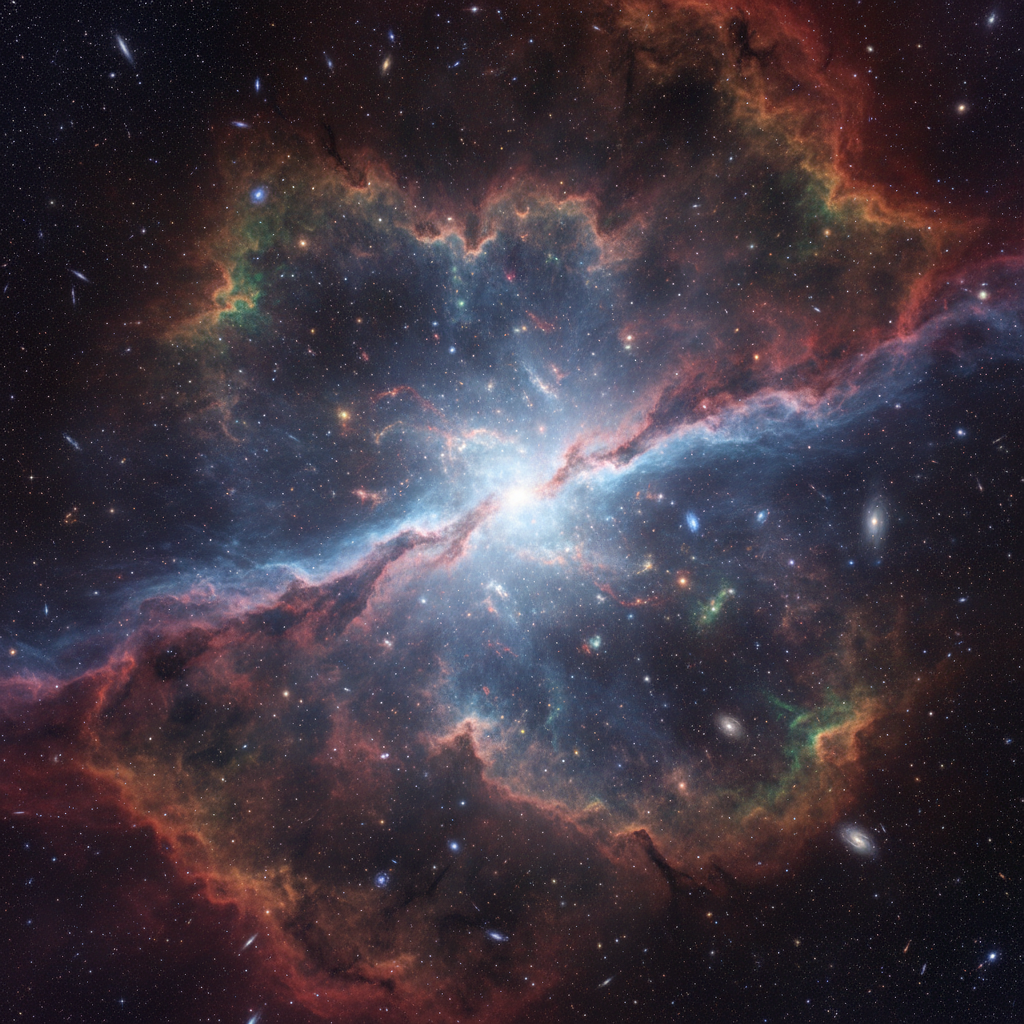
The lab built a spectral decomposition framework — Aperture Spectral Cartography (ASC) — that isolates residuals across eight frequency bands simultaneously, cross-referencing against a database of 2,400 catalogued events. What emerged was not noise. It was a map.
The map suggests that spectral residuals encode local spacetime geometry with sufficient fidelity to distinguish between competing models of quantum gravity — a measurement that was thought to require a detector the size of a solar system.
ASC Framework — Nature Astronomy, Jan 2026
Spectral residual correlation — 8 catalogued sources
The door it opened —
ASC is now being applied to 14 additional black hole systems. Three independent groups have requested the framework. The most consequential finding — a potential signature of Planck-scale geometry — is under peer review.
The question that haunts us now —
Does biological consciousness leave a physical residue in the electromagnetic field?
The question is not new. What is new is the instrument. The same interferometric approach that resolved membrane topology at sub-nanometer scales can, in principle, detect field perturbations at the threshold of biological neural activity — perturbations six orders of magnitude below what existing magnetoencephalography can register.
Aperture's Neuro-Field group began data collection in September 2025. The preliminary signal-to-noise ratio is better than predicted. We cannot yet say what the signal is. We can say that it is structured — periodic, amplitude-modulated, and correlated with reported states of focused attention in 17 of 19 subjects.
The current working hypothesis — one we hold loosely, as all hypotheses should be held — is that the signal represents a previously uncharacterized mode of bioelectromagnetic coherence. If it survives replication, it would require a revision of the standard model of neural field theory.
"We are looking at something that does not yet have a name. The instrument is running. The data is accumulating. What we will find is —"
The real work is one door away.
Full publications, active methodology documentation, open positions, and collaboration enquiries — everything the anteroom pointed toward.